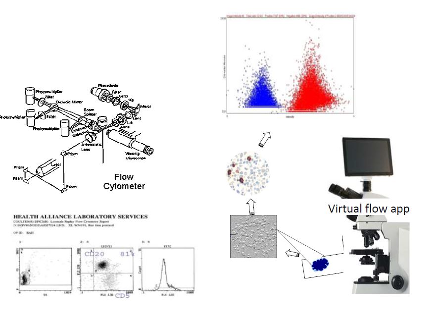
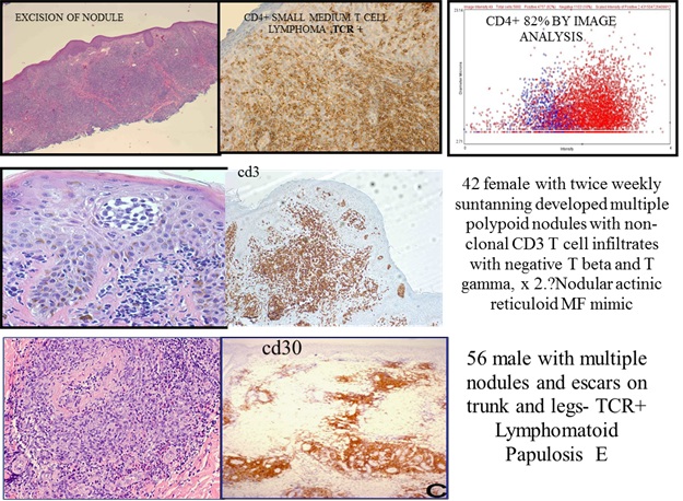
Virtual Flow Cytometry: Conversion of formalin fixed paraffin embedded tissue immunostaining to flow cytometry-like single cell data, (instead of pixel area analysis) with display of data showing population of objects or events that are red positive, blue negative, object distribution according to size in Y axis and intensity in X axis, as a dual parameter dot plot histogram. Throughput of 10,000 cell objects per minute. This technic, which was based on IHCFLOW’s patented color image advanced algorithm, was applied to the real world data to address the question of possible role of biomarkers differential expression in separating subsets of CD30 positive cutaneous lymphoproliferation.
Diagram below shows the similarity of virtual image analysis technic to the cell suspension single cell analysis method used by flow cytometry method.

Details


